- Introduction
- Transport in Plants
- Transpiration
- Translocation of Organic Compounds
- Transport in Animals
- Mammalian Circulatory System
- Structure and Function of Blood
- Immune Responses

Introduction
- Transport is the movement of substances within an organism.
- All living cells require oxygen and food for various metabolic processes.
- These substances must be transported to the cells.
- Metabolic processes in the cells produce excretory products which should be eliminated before they accumulate.
- The excretory products should be transported to sites of excretion.
- Organisms like amoeba are unicellular.
- They have a large surface area to volume ratio.
- The body is in contact with the environment.
- Diffusion is adequate to transport substances across the cell membrane and within the organism.
- Large multi-cellular organisms have complex structure where cells are far from each other hence diffusion alone cannot meet the demand for supply and removal of substances.
- Therefore an elaborate transport system is necessary.
Transport in Plants
- Simple plants such as mosses and liverworts lack specialized transport system.
- Higher plants have specialized transport systems known as the vascular bundle.
- Xylem transports water and mineral salts.
- Phloem transports dissolved food substances like sugars.
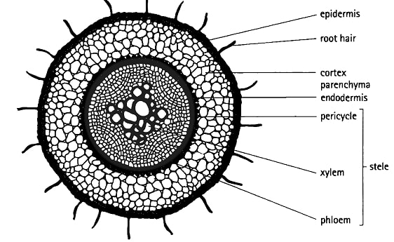
Internal Structure of Roots and Root Hairs
- The main functions of roots are ;
- Anchorage
- absorption.
- storage
- gaseous exchange.
- The outermost layer in a root is the piliferous layer.
- This is a special epidermis of young roots whose cells give rise to root hairs.
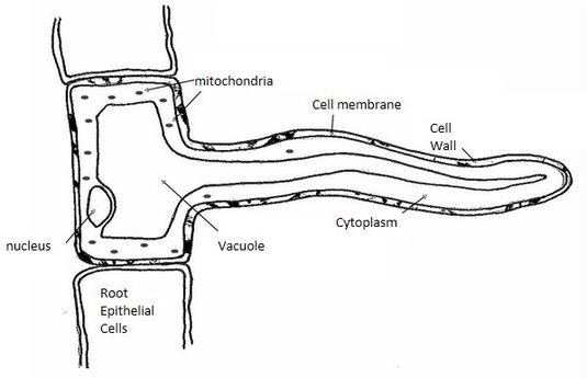
- Root hairs are microscopic outgrowths of epidermal cells.
- They are found just behind the root tip,
- They are one cell thick for efficient absorption of substances.
- They are numerous and elongated providing a large surface area for absorption of water and mineral salts.
- Root hairs penetrate the soil and make close contact with it.
- Below the piliferous layer is the cortex.
- This is made up of loosely packed, thin walled parenchyma cells.
- Water molecules pass through this tissue to reach the vascuiar bundles.
- In some young plant stems, cortex cells contain chloroplasts.
- The endodermis (starch sheath) is a single layer of cells with starch grains.
- The endodermis has a casparian strip which has an impervious deposit controlling the entry of water and mineral salts into xylem vessels.
- Pericyc1e forms a layer next to the endodermis.
- Next to the pericycle is the vascular tissue.
- In the Dicotyledonous root, xylem forms a star shape in the centre, with phloem in between the arms.
- It has no pith. In monocotyledonous root, xylem alternates with phloem and there is a pith in the centre.
Internal Structure of a Root Hair Cell
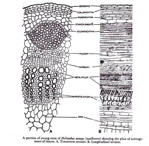
The Stem
- The main functions of the stem are;
- support and exposure of leaves and flowers to the environment,
- conducting water and mineral salts
- conducting manufactured food from leaves to other parts of the plant.
- In monocotyledonous stems, vascular bundles are scattered all over the stem, while in dicotyledonous stems vascular bundles are arranged in a ring.
- Vascular bundles are continuous from root to stems and leaves.
- The epidermis forms a single layer of cells enclosing other tissues.
- The outer walls of the cells have waxy cuticle to prevent excessive loss of water.
- The cortex is a layer next to the epidermis.
- It has collenchyma, parenchyma and schlerenchyma cells.
Collenchyma
- Is next to the epidermis and has thickened walls at the corners which strengthen the stem.
Parenchyma
- Cells are irregular in shape, thin walled and loosely arranged hence creating intercellular spaces filled with air.
- They are packing tissues and food storage areas.
Sclerenchyma
- Cells are closely connected to vascular bundles.
- These cells are thickened by deposition of lignin and they provide support to plants.
Pith
- Is the central region having parenchyma cells.
Absorption of Water and Mineral Salts
Absorption of Water
- Root hair cell has solutes in the vacuole and hence a higher osmotic pressure than the surrounding soil water solution.
- Water moves into the root hair cells by osmosis along a concentration gradient.
- This makes the sap in the root hair cell to have a lower osmotic pressure than the surrounding cells.
- Therefore water moves from root hair cells into the surrounding cortex cells by osmosis.
- The process continues until the water gets into the xylem vessels.
Uptake of Mineral Salts
- If the concentration of mineral salts in solution is greater than its concentration in root hair cell, the mineral salts enter the root hair cell by diffusion.
- If the concentration of mineral salts in the root hair cells is greater than in the soil water, the mineral salts enter the root hairs by active transport.
- Most minerals are absorbed in this way.
- Mineral salts move from cell to cell by active transport until they reach the xylem vessel.
- Once inside the xylem vessels, mineral salts are transported in solution as the water moves up due to root pressure, capillary attraction and cohesion and adhesion forces.
Transpiration
- Transpiration is the process by which plants lose water in the form of water vapour into the atmosphere.
- Water is lost through stomata, cuticle and lenticels.
Stomatal Transpiration:
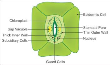
- This accounts for 80-90% of the total transpiration in plants.
- Stomata are found on the leaves.
Cuticular Transpiration:
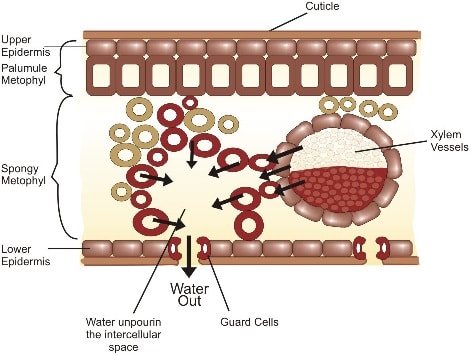
- The cuticle is found on the leaves, and a little water is lost through it.
- Plants with thick cuticles do not lose water through the cuticle.
Lenticular Transpiration
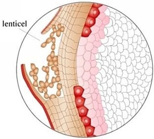
- Is loss' of water through lenticels.
- These are found on stems of woody plants.
- Water lost through the stomata and cuticle by evaporation leads to evaporation of water from surfaces of mesophyll cells .
- The mesophyll cells draw water from the xylem vessels by osmosis.
- The xylem in the leaf is continuous with xylem in the stem and root.
Structure and Function of Xylem
- Movement of water is through the xylem.
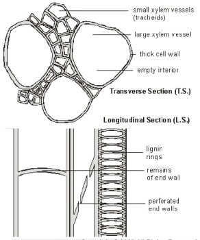
- Xylem tissue is made up of vessels and tracheids.
Xylem Vessels
- Xylem vessels are formed from cells that are elongated along the vertical axis and arranged end to end.
- During development, the cross walls and organelles disappear and a continuous tube is formed.
- The cells are dead and their walls are strengthened by deposition of lignin.
- The lignin has been deposited in various ways.
- This results in different types of thickening
- Annular.
- Simple spiral.
- Double spiral.
- Reticulate.
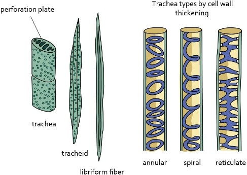
- The bordered pits are areas without lignin on xylem vessels and allow passage of water in and out of the lumen to neighbouring cells.
Tracheids
- Tracheids have cross-walls that are perforated.
- Their walls are deposited with lignin.
- Unlike the xylem vessels, their end walls are tapering or chisel-shaped.
- Their lumen is narrower.
- Besides transport of water, xylem has another function of strengthening the plant which is provided by xylem fibres and xylem parenchyma.
Xylem Fibres ;
- Are cells that are strengthened with lignin.
- They form wood.
Xylem Parenchyma:
- These are cells found between vessels.
- They form the packing tissue.
Forces Involved in Transportation of Water and Mineral Salts
Transpiration Pull
- As water vaporises from spongy mesophyll cells into sub-stomatal air spaces, the cell sap of mesophyll cells develop a higher osmotic pressure than adjacent cells.
- Water is then drawn into mesophyll cells by osmosis from adjacent cells and finally from xylem vessels.
- A force is created in the leaves which pulls water from xylem vessels in the stem and root.
- This force is called transpiration pull.
Cohesion and Adhesion:
- The attraction between water molecules is called cohesion.
- The attraction between water molecules and the walls of xylem vessels is called adhesion.
- The forces of cohesion and adhesion maintain a continuous flow of water in the xylem from the root to the leaves.
Capillarity:
- Is the ability of water to rise in fine capillary tubes due to surface tension.
- Xylem vessels are narrow, so water moves through them by capillarity.
Root Pressure:
- If the stem of a plant is cut above the ground level, it is observed that cell sap continues to come out of the cut surface.
- This shows that there is a force in the roots that pushes water up to the stem.
- This force is known as root pressure.
Importance of Transpiration
- Transpiration leads to excessive loss of water if unchecked.
Some beneficial effects are:
- Replacement of water lost during the process.
- Movement of water up the plant is by continuous absorption of water from the soil.
- Mineral salts are transported up the plant.
- Transpiration ensures cooling of the plant in hot weather.
- Excessive loss of water leads to wilting' and eventually death if water is not available in the soil.
Factors Affecting Transpiration
The factors that affect transpiration are grouped into two. i.e. environmental and structural.
Environmental Factors
Temperature
- High temperature increases the internal temperature of the leaf which in turn increases kinetic energy of water molecules which increases evaporation.
- High temperatures dry the air around the leaf surface maintaining a high concentration gradient.
- More water vapour is therefore lost from the leaf to the air.
Humidity
- The higher the humidity of the air around the leaf, the lower the rate of transpiration.
- The humidity difference between the inside of the leaf and the outside is called the saturation deficit.
- In dry atmosphere, the saturation deficit is high.
- At such times, transpiration rate is high.
Wind
- Wind carries away water vapour as fast as it diffuses out of the leaves.
- This prevents the air around the leaves from becoming saturated with vapour.
- On a windy day, the rate of transpiration is high.
Light Intensity
- When light intensity is high; more stomata open hence high rate of transpiration.
Atmospheric Pressure
- The lower the atmospheric pressure the higher the kinetic energy of water molecules hence more evaporation.
- Most of the plants at higher altitudes where atmospheric pressure is very low have adaptations to prevent excessive water-loss.
Availability of Water
- The more water there is in the soil, the more is absorbed by the plant and hence a lot of water is lost by transpiration.
Structural Factors
Cuticle
- Plants growing in arid or semi-arid areas have leaves covered with a thick waxy cuticle.
Stomata
- The more the stomata, the higher the rate of transpiration.
- Xerophytes have few stomata which reduce water-loss.
- Some have sunken stomata which reduces the rate of transpiration as the water vapour accumulates in the pits.
- Others have stomata on the lower leaf surface hence reducing the rate of water-loss.
- Some plants have reversed stomatal rhythm whereby stomata close during the day and open at night.
- This helps to reduce water-loss.
Leaf Size and Shape
- Plants in wet areas have large surface area for transpiration.
- Xerophytes have small narrow leaves to reduce water-loss.
- The photometer can be used to determine transpiration in different environmental conditions.
Translocation of Organic Compounds
- Translocation of soluble organic products of photosynthesis within a plant is called translocation.
- It occurs in phloem in sieve tubes.
- Substances translocated include glucose, amino acids, vitamins.
- These are translocated to the growing regions like stem, root apex, storage organs e.g. corms, bulbs and secretory organs such as nectar glands.
Phloem
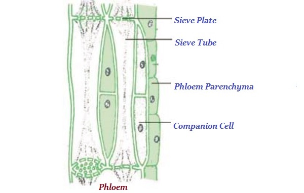
phloem is made up of;
- sieve tubes,
- companion cells
- parenchyma, a packing tissue
- schlerenchyma, a strengthening tissue
Sieve Tubes
- These are elongated cells arranged end to end along the vertical axis.
- The cross walls are perforated by many pores to make a sieve plate.
- Most organelles disappear and those that remain are pushed to the sides of the sieve tube.
- Cytoplasmic strands pass through the pores in the plate into adjacent cells.
- Food substances are translocated through cytoplasmic strands.
Companion Cells
- Companion cells are small cells with large nuclei and many mitochondria.
- They are found alongside each sieve element.
- The companion cell is connected to the tube through plasmodesmata.
- The mitochondria generate energy required for translocation.
Phloem Parenchyma
- These are parenchyma cells between sieve elements.
- They act as packing tissue.
Transport in Animals
The Circulatory System
- Large and complex animals have circulatory systems that consist of tubes, a transport fluid and a means of pumping the fluid.
- Blood is the transport fluid which contains dissolved substances and cells.
- The tubes are blood vessels through which dissolved substances are circulated around the body.
- The heart is the pumping organ which keeps the blood in circulation.
The types of circulatory system exist in animals: open and closed.
In an open circulatory system;
- The heart pumps blood into vessels which open into body spaces known as haemocoel.
- Blood comes into contact with tissues.
A closed circulatory system;
- Found in vertebrates and annelids where the blood is confined within blood vessels and does not come into direct contact with tissues.
Transport in Insects
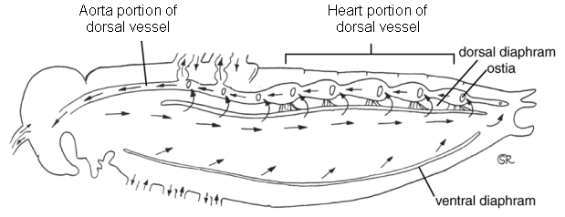
- In an insect, there is a tubular heart just above the alimentary canal.
- This heart is suspended in a pericardial cavity by ligaments.
- The heart has five chambers and extends along the thorax and abdomen.
- Blood is pumped forwards into the aorta by waves of contractions in the heart.
- It enters the haemocoel and flows towards the posterior.
- The blood flows back into the heart through openings in each chamber called ostia.
- The ostia have valves which prevent the backflow of blood.
- Blood is not used as a medium for transport of oxygen in insects.
- This is because oxygen is supplied directly to the tissues by the tracheal system.
- The main functions of blood in an insect are to transport nutrients, excretory products and hormones.
Mammalian Circulatory System
- Mammals have a closed circulatory system where a powerful heart pumps blood into arteries.
- The arteries divide into smaller vessels called arterioles.
- Each arteriole divides to form a network of capillaries inside the tissues.
- The capillaries eventually re-unite to form venules, which form larger vessels called veins.
- The veins take the blood back to the heart.
- Blood from the heart goes through the pulmonary artery to the lungs and then back to the heart through pulmonary vein.
- This circulation is called pulmonary circulation.
- Oxygenated blood leaves the heart through the aorta and goes to all the tissues of the body.
- From the tissues, deoxygenated blood flows back to the heart through the vena cava.
- This circulation is called systemic circulation.
- In each complete circulation, the blood flows into the heart twice.
- This is called double circulation.
- Some other animals like fish have a single circulation.
- Blood flows only once through the heart for every complete circuit.
Structure and Function of the Heart
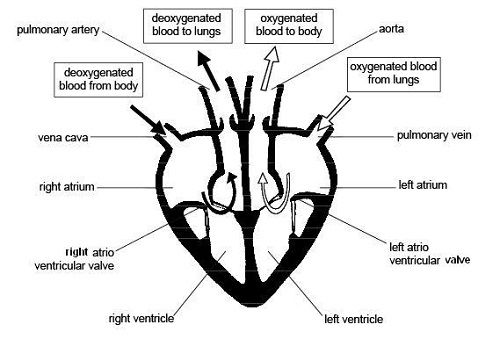
- The heart has four chambers:
- Two artria (auricles) and two ventricles.
- The left and right side of the heart are separated by a muscle wall (septum) so that oxygenated and deoxygenated blood does not mix.
- Deoxygenated blood from the rest of the body enters the heart through the vena cava.
- Blood enters the right atrium, then through tricuspid valve into right ventricle.
- Then via semi-lunar valve to the pulmonary artery to the lungs.
- Oxygenated blood from the lungs enters the heart through pulmonary vein.
- It enters the left atrium of the heart, then through bicuspid valve into left ventricle.
- Then via semi-lunar valves to aorta which takes oxygenated blood round the body.
- A branch of the aorta called coronary artery supplies blood to the heart muscle.
- The coronary vein carries blood from the heart muscle to the pulmonary artery which then takes it to the lungs for oxygenation.
Pumping Mechanism of the Heart
- The heart undergoes contraction (systole) and relaxation (diastole).
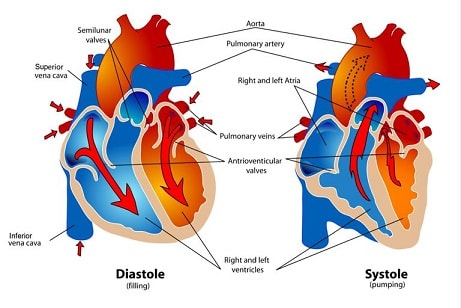
Systole
- When the ventricular muscles contract, the cuspid valves (tricuspid and bicuspid) close preventing backflow of blood into auricles.
- The volume of the ventricles decreases while pressure increases.
- This forces blood out of the heart to the lungs through semi-lunar valves and pulmonary artery, and to the body tissues via semi-lunar valve and aorta respectively.
- At the same time the atria are filled with blood.
- The left ventricle has thicker muscles than the right ventricle, and pumps blood for a longer distance to the tissues.
Diastole
- When ventricular muscles relax, the volume of each ventricle increases while pressure decreases.
- Contractions of atria force the bicuspid and tricuspid valves to open allowing deoxygenated blood from right atrium into right ventricle which oxygenated blood flows from left atrium into the left ventricle.
- Semi-lunar valves close preventing the backflow of blood into ventricles.
- The slight contractions of atria force the , blood flow into ventricles.
The Heartbeat
- The heart is capable of contracting and relaxing rhythmically without fatigue due to its special muscles called cardiac muscles.
- The rhythmic contraction of the heart arise from within the heart muscles without nervous stimulation.
- The contraction is said to be myogenic.
- The heartbeat is initiated by the pacemaker or sino-artrio-node (SAN) which is located in the right atrium.
- The wave of excitation spreads over the walls of atria.
- It is picked by the artrio-ventricular node which is located at the junction of the atria and ventricles, from where the purkinje tissue spreads the wave to the walls of the ventricles.
- The heart contracts and relaxes rhythmically at an average rate of 72 times per minute.
- The rate of the heartbeat is increased by the sympathetic nerve, while it is slowed down by the vagus nerve.
- Heartbeat is also affected by hormones e.g. adrenaline raises the heartbeat.
Structure and Function of Arteries, Capillaries and Veins
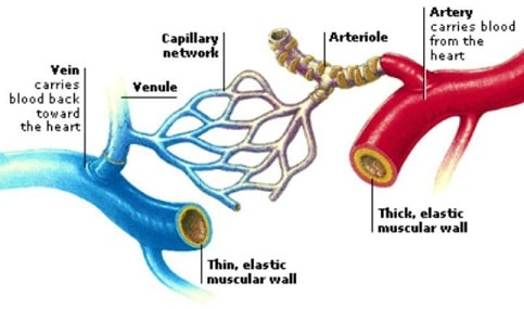
Arteries
- Arteries carry blood away from the heart.
- They carry oxygenated blood except pulmonary artery which carries deoxygenated blood to the lungs.
- Arteries have a thick, muscular wall, which has elastic and collagen fibres that resist the pressure of the blood flowing in them.
- The high pressure is due to the pumping action of the heart.
- The pressure in the arteries originate from the pumping action of the heart.
- The pulse or number of times the heart beats per minute can be detected by applying pressure on an artery next to the bone. e.g. by placing the finger/thumb on the wrist.
- The innermost layer of the artery is called endothelium which is smooth.
- It offers least possible resistance to blood flow.
- Have a narrow lumen.
- The aorta forms branches which supply blood to all parts of the body.
- These arteries divide into arterioles which further divide to form capillaries.
Capillaries
- Capillaries are small vessels whose walls are made of endothelium which is one cell thick.
- This provides a short distance for exchange of substances.
- Capillaries penetrate tissues,
- The lumen is narrow therefore blood flowing in capillaries is under high pressure.
- Pressure forces water and dissolved substances out of the blood to form tissue fluid.
- Exchange of substances occurs between cells and tissue fluid.
- Part of the tissue fluid pass back into capillaries at the venule end.
- Excess fluid drains into small channels called lymph capillaries which empty their contents into lymphatic vessels.
- Capillaries join to form larger vessels called venules which in turn join to form veins which transport blood back to the heart.
Veins
- Veins carry deoxygenated blood from the tissues to the heart (except pulmonary vein which carries oxygenated blood from the lungs to the heart).
- Veins have a wider lumen than arteries.
- Their walls are thinner than those of arteries.
- Blood pressure in the veins is low.
- Forward flow of blood in veins is assisted by contraction of skeletal muscles, hence the need for exercise.
- Veins have valves along their length to prevent backflow of blood.
- This ensures that blood flows towards the heart.
- The way the valves work can be demonstrated on the arm.
- By pressing on one vein with two fingers, leaving one and pushing blood toward the heart then releasing the latter finger, it can be observed that the part in between is left with the vein not visible.
- This is because blood does not flow back towards the first finger.
Diseases and Defects of Circulatory System
Thrombosis
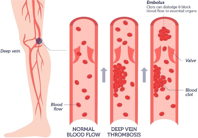
- Formation of a clot in the blood vessels is called thrombosis.
- Coronary thrombosis is the most common.
- It is caused by blockage of coronary artery which supplies blood to the heart.
- Blockage may be due to artery becoming fibrous or accumulation of fatty material on the artery walls.
- Narrow coronary artery results in less blood reaching the heart muscles.
- A serious blockage can result in heart attack which can be fatal.
- Heavy intake of fat, alcohol, being overweight and emotional stress can cause coronary thrombosis.
- A blockage in the brain can lead to a stroke causing paralysis of part of the body, coma or even death.
- A healthy lifestyle, avoiding a lot of fat in meals and avoiding alcohol can control the disease.
Arteriosclerosis
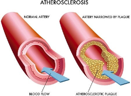
- This condition results from the inner walls having materials being deposited there or growth of fibrous connective tissue.
- This leads to thickening of the wall of the artery and loss of elasticity.
- Normal blood flow is hindered.
- Arteriosclerosis can lead to thrombosis or hypertension.
- A person with hypertension which is also called high blood pressure has his/her blood being pumped more forcefully through the narrow vessels.
- This puts stress on the walls of the heart and arteries.
- Regular exercise, healthy diet and avoiding smoking can help maintain normal blood pressure.
Varicose Veins
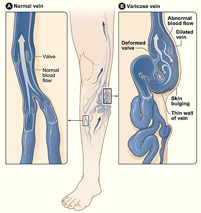
- Superficial veins especially at the back of the legs become swollen and flabby due to some valves failing to function properly.
- This results to retention of tissue fluid.
- Regular physical exercise will prevent this condition.
- Repair of valves through surgery can also be done.
- Wearing surgical stockings may ease a mild occurence.
Structure and Function of Blood
Composition of Blood
- The mammalian blood is made up of a fluid medium called plasma with substances dissolved in it.
- Cellular components suspended in plasma include;
- erythrocytes (red blood cells),
- leucocytes (white blood cells)
- thrombocytes (platelets)
- blood proteins.
Plasma
- This is a pale yellow fluid consisting of 90% water.
- There are dissolved substances which include;
- glucose, amino acids, lipids, salts,
- hormones, urea, fibrinogen, albumen,
- antibodies, some enzymes suspended cells.
- Serum is blood from which fibrinogen and cells have been removed.
The functions of plasma include:
- Transport of red blood cells which carry oxygen.
- Transport dissolved food substances round the body.
- Transport metabolic wastes like nitrogenous wastes and carbon (IV) oxide in solution about 85% of the carbon (IV) oxide is carried in form of hydrogen carbonates.
- Transport hormones from sites of production to target organs.
- Regulation of pH of body fluids.
- Distributes heat round the body hence regulate body temperature.
Erythrocytes (Red Blood Cells)
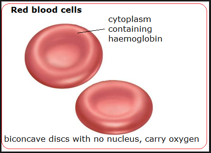
- In humans these cells are circular biconcave discs without nuclei.
- Absence of nucleus leaves room for more haemoglobin to be packed in the cell to enable it to carry more oxygen.
- Haemoglobin contained in red blood cells is responsible for the transport of oxygen.
- Haemoglobin + Oxygen =oxyhaemoglobin
(Hb) + (4O2) __ (HbOg) - Oxygen is carried in form of oxyhaemoglobin.
- Haemoglobin readily picks up oxygen in the lungs where concentration of oxygen is high.
- In the tissues, the oxyhaemoglobin breaks down (dissociates) easily into haemoglobin and oxygen.
- Oxygen diffuses out of the red blood cells into the tissues.
- Haemoglobin is then free to pick up more oxygen molecules.
- The biconcave shape increases their surface area over which gaseous exchange takes place.
- Due to their ability, they are able to change their shape to enable themselves squeeze inside the narrow capillaries.
- CO2+ H2O carbonic anhydrase
- There are about five million red blood cells per cubic millimetre of blood.
- They are made in the bone marrow of the short bones like sternum, ribs and vertebrae.
- In the embryo they are made in the liver and spleen.
- Erythrocytes have a life span of about three to four months after which they are destroyed in the liver and spleen.
- Also in the red blood cells is carbonic anhydrase which assists in the transport of carbon (IV) oxide.
Leucocytes (White Blood Cells)
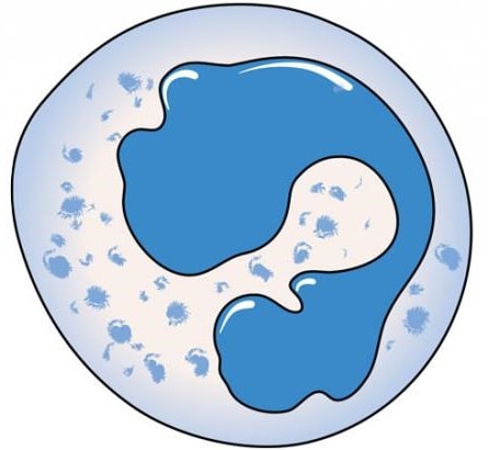
- These white blood cells have a nucleus.
- They are divided into two:
- Granulocytes (also phagocytes or polymorphs)
- Agranulocytes
- White blood cells defend the body against disease.
- Neutrophils form 70% of the granulocytes.
- Others are eosinophils and basophils.
- About 24% agronulocytes are called lymphocytes, while 4% agranulocytes are monocytes.
- The leucocytes are capable of amoebic movement.
- They squeeze between the cells of the capillary wall to enter the intercellular spaces.
- They engulf and digest disease causing organisms (pathogens) by phagocytosis.
- Some white blood cells may die in the process of phagocytosis.
- The dead phagocytes, dead organisms and damaged tissues form pus.
- Lymphocytes produce antibodies which inactivate antigens.
Antibodies include: - Antitoxins which neutralise toxins.
- Agglutinins cause bacteria to clump together and they die.
- Lysins digest cell membranes of microorganisms.
- Opsonins adhere to outer walls of microorganisms making it easier for phagocytes to ingest them.
- Lymphocytes' are made in the thymus gland and lymph nodes.
- There are about 7,000 leucocytes per cubic millimetre of blood.
Platelets (Thrombocytes)
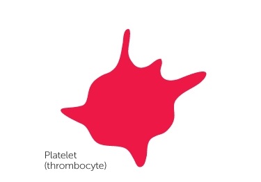
- Platelets are small irregularly shaped cells formed from large bone marrow cells called megakaryocytes.
- There are about 250,000 platelets per cubic millimetre of blood.
- They initiate the process of blood clotting.
- The process of clotting involves a series of complex reactions whereby fibrinogen is converted into a fibrin clot.
- When blood vessels are injured platelets are exposed to air and they release thromboplastin (thrombokinase) which initiates the blood clotting process.
- Thromboplastin neutralises heparin the anti-clotting factor in blood and activates prothrombin to thrombin.
- The process requires calcium ions and vitamin K.
- Thrombin activates the conversion of fibrinogen to fibrin which forms a meshwork of fibres on the cut surface to trap red blood cells to form a clot.
- The clot forms a scab that stops bleeding and protects the damaged tissues from entry of micro-organisms.
- Blood clotting reduces loss of blood when blood vessels are injured.
- Excessive loss of blood leads to anaemia and dehydration.
- Mineral salts lost in blood leads to osmotic imbalance in the body.
- This can be corrected through blood transfusion and intravenous fluid.
ABO Blood Groups
- There are four types of blood groups in human beings: A, B, AB and O.
- These are based on types of proteins on the cell membrane of red blood cells.
- There are two types of proteins denoted by the letters A and B which are antigens.
- In the plasma are antibodies specific to these antigens denoted as a and b.
- A person of blood group A has A antigens on the red blood cells and b antibodies in plasma.
- A person of blood group B has B antigens on red blood cells and a antibodies in plasma.
- A person of blood group AB has A and B antigens on red blood cells and no antibodies in plasma .
- A person of blood group a has no antigens on red blood cells and a and b antibodies in plasma.
Blood groups
Blood
GroupsAntigen Antibodies A A b B B a AB A and B None O None a and b
Blood Transfusion
Blood transfusion is the transfer of blood from a donor to the circulatory system of the recipient.
A recipient will receive blood from a donor if the recipient has no corresponding antibodies to the donor's antigens.
If the donor's blood and the recipient's blood are not compatible, agglutination occurs whereby red blood cells clump together.
Blood Typing
- A person of blood group 0 can donate blood to a person of any other blood group.
- A person of blood group 0 is called a universal donor.
- A person of blood group AB can receive blood from any other group.
- A person with blood group AB is called a universal recipient.
- A person of blood group A can only donate blood to another person with blood group A or a person with blood group AB.
- A person of blood group B can only donate blood to somebody with blood group B or a person with blood group AB.
- A person with blood group AB can only donate blood to a person with blood group AB.
- Blood screening has become a very important step in controlling HIV/AIDS.
- It is therefore important to properly screen blood before any transfusion is done.
Rhesus Factor
- The Rhesus factor is present in individuals with the Rhesus antigen in their red blood cells.
- Such individuals are said to be Rhesus positive (Rh+), while those without the antigen are Rhesus negative (Rh-).
- If blood from an Rh+ individual is introduced into a person who is Rh- , the latter develops antibodies against the Rhesus factor.
- There may not be any reaction after this transfusion.
- However a subsequent transfusion with Rh+ blood causes a severe reaction, and agglutination occurs i.e. clumping of red blood cells.
- The clump can block the flow of blood, and cause death.
- Erythroblastosis foetalis (haemolytic disease of the newborn) results when an Rh- mother carries an Rh+ foetus.
- This arises when the father is Rh+.
- During the latter stage of pregnancy, fragments of Rhesus positive red blood cells of the foetus may enter mother's circulation.
- These cause the mother to produce Rhesus antibodies which can pass across the placenta to the foetus and destroy foetal red blood cells.
- During the first pregnancy, enough antibodies are not formed to affect the foetus.
- Subsequent pregnancies result in rapid production of Rhesus antibodies by the mother.
- These destroy the red blood cells of the foetus, the condition called haemolytic disease of the newborn.
- The baby is born anaemic and with yellow eyes (jaundiced).
- The condition can be corrected by a complete replacement of baby's blood with safe healthy blood.
Lymphatic System
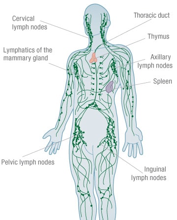
- The lymphatic system consists of lymph vessels.
- Lymph vessels have valves to ensure unidirectional movement of lymph.
- Lymph is excess tissue fluid i.e. blood minus blood cells and plasma proteins.
- Flow of lymph is assisted by breathing and muscular contractions.
- Swellings called lymph glands occur at certain points along the lymph vessels.
- Lymph glands are oval bodies consisting of connective tissues and lymph spaces.
- The lymph spaces contain lymphocytes which are phagocytic.
- Lymph has the same composition as blood except that it does not contain red blood cells and plasma proteins.
- Lymph is excess tissue fluid.
- Excess tissue fluid is drained into lymph vessels by hydrostatic pressure.
- The lymph vessels unite to form major lymphatic system.
- The main lymph vessels empty the contents into sub-clavian veins which take it to the heart.
Immune Responses
- Immune response is the production of antibodies in response to antigens.
- An antigen is any foreign material or organism that is introduced into the body and causes the production of antibodies.
- Antigens are protein in nature.
- An antibody is a protein whose structure is complementary to the antigen.
- This means that a specific antibody deals with a specific antigen to make it harmless.
- When harmful organisms or proteins invade the body, lymphocytes produce complementary antibodies, while bone marrow and thymus gland produce more phagocytes and lymphocytes respectively.
Types of Immunity
- There are two types of immunity; natural and artificial.
- Natural Immunity is also called innate immunity. It is inherited from parent to offspring.
- Artificial Immunity can be natural or induced.
- attacked by diseases like chicken pox, measles and mumps, those who recover from these diseases develop resistance to any subsequent infections of the same diseases. This is natural acquired immunity.
Artificial Acquired Immunity:
- When attenuated (weakened) or dead microorganisms are introduced into a healthy person.
- The lymphocytes synthesis the antibodies which are released into the lymph and eventually reach the blood.
- The antibodies destroy the invading organisms.
- The body retains 'memory' of the structure of antigen.
- Rapid response is ensured in subsequent infections.
- Vaccines generally contain attenuated disease causing organisms.
Artificial Passive Acquired Immunity:
- Serum containing antibodies is obtained from another organism, and confers immunity for a short duration.
- Such immunity is said to be passive because the body is not activated to produce the antibodies.
Importance of Vaccination
- A vaccine is made of attenuated, dead or nonvirulent micro-organism that stimulate cells in the immune system to recognise and attack disease causing agent through production of antibodies.
- Vaccination protects individuals from infections of many diseases like smallpox, tuberculosis and poliomyelitis.
- Diseases like smallpox, tuberculosis and tetanus were killer diseases but this is no longer the case.
- Diphtheria Pertussis Tetanus (DPT) vaccine protects children against diphtheria, whooping cough and tetanus.
- Bacille Calmette Guerin (BCG) vaccine is injected at birth to children to protect them against tuberculosis.
- Measles used to be a killer disease but today, a vaccine injected into children at the age of nine months prevents it.
- At birth children are given an inoculation through the mouth of the poliomyelitis vaccine.
Allergic Reactions
- An allergy is a hypersensitive reaction to an antigen by the body.
- The antibody reacts with the antigen violently.
- People with allergies are oversensitive to foreign materials like dust, pollen grains, some foods, some drugs and some air pollutants.
- Allergic reactions lead to production of histamine by the body.
- Histamine causes swelling and pain.
- Allergic reactions can be controlled by avoiding the allergen and administration of anti-histamine drugs.
Join our whatsapp group for latest updates
Tap Here to Download for 50/-
Get on WhatsApp for 50/-
Download TRANSPORT IN PLANTS AND ANIMALS - Form 2 Biology Notes.
Tap Here to Download for 50/-
Get on WhatsApp for 50/-
Why download?
- ✔ To read offline at any time.
- ✔ To Print at your convenience
- ✔ Share Easily with Friends / Students